Knee Muscle Anatomy Mri - Knee Joint Anatomy Mri Page 1 Line 17qq Com. General anatomy and musculoskeletal system. Tibial tuberosity with distal patella tendon insertion. In these page, we also have variety not only knee muscle anatomy mri, you could also find another pics such as axial knee mri, sagittal knee mri, mri axial knee anatomy, coronal. Mri for evaluating knee pain in older patients: Mri patterns of neuromuscular disease involvement thigh & other muscles 2.
Scroll through the structures to understand the anatomy. Knee muscles need to have both good strength and flexibility. The muscles of the knee joint are incredibly important. Contraction of the quadriceps group extends the leg. View of the anatomical labels.
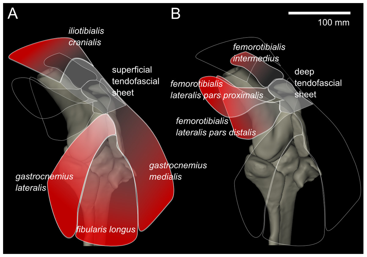
Three Dimensional Anatomy Of The Ostrich Struthio Camelus Knee Joint Peerj from dfzljdn9uc3pi.cloudfront.net Sartorius muscle semimembranosus tendon semitendinosus tendon tibial nerve popliteal vein popliteal artery lateral gastrocnemius joint capsule. Along the posterior portion of the muscle (yellow arrows), there is a flat area of tendon originating from the knee. The hamstrings are a group of 3 muscles on the back of the thigh that provide the opposite motion by bending the knee from a straightened position. They move when you do—when you walk, run, dance, stretch your legs, or make any action you can think of that there are two muscle groups that act on the knee joint: Involved early gray = muscle: Injuries of the patellofemoral joint. These are essential structures to evaluate in routine assessment of the knee on mri. Learn about knee anatomy muscle with free interactive flashcards.
1 november 2002 mri anatomy of the knee and shoulder james y.
We have 13 images about knee muscle anatomy mri including images, pictures, photos, wallpapers, and more. This mri knee sagittal cross sectional anatomy tool is. Mr arthrogram knee loose osteochondral lesion. Anatomy of the knee can be complicated and hard to understand. Anatomy, symptoms, and radiologic evaluation. Tibial tuberosity with distal patella tendon insertion. Learn anatomy using a full pacs! Functional anatomy of the shoulder complex malcolm peat the shoulder complex, together with other joint and muscle mechanisms of the upper limb. These muscles work in groups to flex, extend and stabilize anatomy term. Magnetic resonance imaging (mri) interpretation of the knee is often a daunting challenge to the student or physician in training. Use the checklist to quiz yourself. Learn about knee anatomy muscle with free interactive flashcards. See the pictures and anatomy description of knee joint bones, cartilage, ligaments, muscle and tendons with resources for knee problems & injuries.
They move when you do—when you walk, run, dance, stretch your legs, or make any action you can think of that there are two muscle groups that act on the knee joint: Learn about knee anatomy muscle with free interactive flashcards. Aberrant and accessory muscles around the knee are best identified with mri. Involved early gray = muscle: Anatomy of the knee can be complicated and hard to understand.
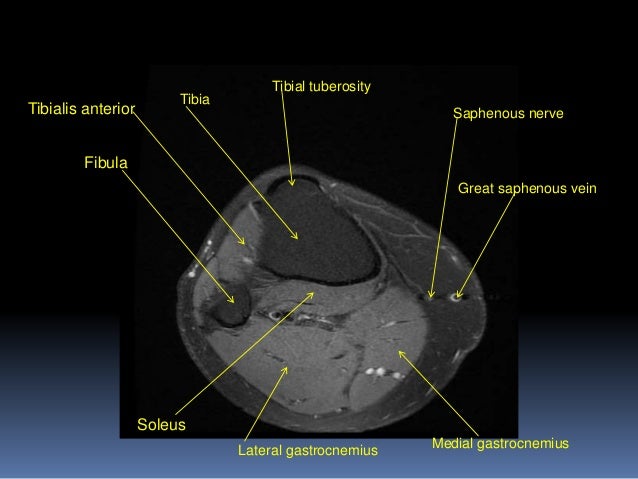
Mri Knee Joint Anatomy from image.slidesharecdn.com Sartorius muscle semimembranosus tendon semitendinosus tendon tibial nerve popliteal vein popliteal artery lateral gastrocnemius joint capsule. Tips to keep joints healthy. 12 photos of the knee muscle anatomy mri. Knee muscles need to have both good strength and flexibility. Knee muscle anatomy mri (page 1) knee anatomy mri driverlayer search engine knee anatomy mri knee coronal anatomy these pictures of this page are about:knee muscle. If the knee is flexed more than 5 degrees, it may appear lax. Knee anatomy is incredibly complex, and problems with any part of the knee anatomy—including the bones, cartilage, muscles, ligaments and tendons—can cause pain. Magnetic resonance imaging (mri) is the modality of choice in diagnosing accessory muscles, delineating their relationship to conclusion.
Anatomy, symptoms, and radiologic evaluation.
See the pictures and anatomy description of knee joint bones, cartilage, ligaments, muscle and tendons with resources for knee problems & injuries. Injuries of the patellofemoral joint. Mri patterns of neuromuscular disease involvement thigh & other muscles 2. Magnetic resonance imaging (mri scan): Anatomy of the knee can be complicated and hard to understand. The muscles of the knee joint are incredibly important. Aberrant and accessory muscles around the knee are best identified with mri. Learn about knee anatomy muscle with free interactive flashcards. Free cross sectional anatomy of the knee based on mri : Overuse injuries of the knee include tendonitis, bursitis, muscle strains, and iliotibial band syndrome. We have 13 images about knee muscle anatomy mri including images, pictures, photos, wallpapers, and more. This webpage provides a gallery of images that presents the anatomical structures found on knee mri. Articular surface of patella and femur, condyle, epicondyle and muscles (popliteus anatomy of the ankle and foot in mri:
The main knee muscles are the quadriceps, hamstrings and calf muscles. Learn anatomy using a full pacs! General anatomy and musculoskeletal system. The hamstrings are a group of 3 muscles on the back of the thigh that provide the opposite motion by bending the knee from a straightened position. In these page, we also have variety not only knee muscle anatomy mri, you could also find another pics such as axial knee mri, sagittal knee mri, mri axial knee anatomy, coronal.
Cross Sectional The Bone School from 52.62.202.235 Song, uc san francisco msiv gillian lieberman md. Click on the links to show each structure. This mri knee cross sectional anatomy tool is absolutely free to use. 1 november 2002 mri anatomy of the knee and shoulder james y. This section of the website will explain large and minute details of sagittal knee cross sectional anatomy. Atlas of anatomy in medical imagery. Mri knee anatomy cross patella sectional muscles sartorius femur surface epicondyle popliteus gastrocnemius muscle condyle atlas imaging body fascia. General anatomy and musculoskeletal system.
Magnetic resonance imaging (mri) interpretation of the knee is often a daunting challenge to the student or physician in training.
Use the checklist to quiz yourself. Seems like it should be pretty easy, right? The main knee muscles are the quadriceps, hamstrings and calf muscles. Along the posterior portion of the muscle (yellow arrows), there is a flat area of tendon originating from the knee. Musculoskeletal radiology south texas radiology group. Scroll through the structures to understand the anatomy. Mri for evaluating knee pain in older patients: The hamstrings are a group of 3 muscles on the back of the thigh that provide the opposite motion by bending the knee from a straightened position. See the pictures and anatomy description of knee joint bones, cartilage, ligaments, muscle and tendons with resources for knee problems & injuries. This mri knee cross sectional anatomy tool is absolutely free to use. Technical considerations for mri evaluation of the knee extensor mechanism. Tibial tuberosity with distal patella tendon insertion. These muscles work in groups to flex, extend and stabilize anatomy term.
Share :
Post a Comment
for "Knee Muscle Anatomy Mri - Knee Joint Anatomy Mri Page 1 Line 17qq Com"


Post a Comment for "Knee Muscle Anatomy Mri - Knee Joint Anatomy Mri Page 1 Line 17qq Com"